ORAL-MAXILOFACIAL SURGERY
Dental extraction is that part of craniofacial or oral surgery which involves the practice of tooth (or a part of one) extraction from the bone that holds it, by means of adequate techniques and instruments.
Dental extraction is the basis of Oral Surgery. It involves simple extraction, as well as surgical: of a root-rest, teeth with position anomalies or a more or less ectopic (out of place) location.

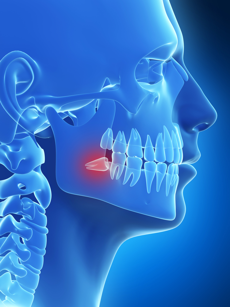
The back teeth or wisdom teeth
They are the last teeth to appear and to develop in the mouth.
Frequently the mouth does not have enough room to accommodate the wisdom teeth. When this occurs, the teeth are retained (trapped by other teeth or by the very bone, underneath the gingiva tissue). If the teeth are retained, they cause pain and swelling in the area.
Wisdom teeth that come out partially or rotated can be the source of various kinds of discomfort;
Dental epulis or cracked epulis
It tends to be more frequent in patients with edentulism (no teeth) in the total upper part and wearers of conventional prosthesis.
It is more frequent in older people. The dental prosthesis with its movement produces an increase of the reabsorption of the alveolar ridge bone, which involves the embedding of the edge of the flap of the prosthesis. This produces an irritation of the underlying mucous membrane which manifests itself in folds, giving name to this entity.
Periapical Surgery
Some patients have lesions in the maxillary bone or jaw, around one or several roots, that destroy the supporting bone of the tooth. They are responsible for pain and infection. These lesions are called granulomas or periapical cysts and their origin is from chronic dental infection.
When these lesions are a small size, less than 1 centimetre, the treatment is carried out through the tooth root canal. Normally, the root canal solves the problem, although not in all cases.
When the root canal does not allow healing of the lesion, a repetition of the root canal treatment is indicated. If this does not control the evolution of the lesion, periapical surgery would be indicated.
Periapical Surgery consists of the surgical removal of the lesion found at the end of the root of the tooth, along with the section of the final part of the root
(about 3 mm). Normally, it is accompanied by the performance of a small preparation at the end of the sectioned root and a retrograde sealing of it with amalgam without zinc (Zn) or with special cement, so that it is well sealed and no germs get in.
Maxillary-facial Surgery
 INCLUDED TEETH
The included teeth are teeth that have not come out during their normal period and remain, partially or totally, inside the bone.
Any tooth can suffer this process of inclusion, but it tends to affect, above all, the back upper and lower (wisdom teeth) and the upper canine teeth. This is due to the fact that these are the last ones to come out and, therefore, have more problems because they have less room.
In these cases we must rule out any type of pathology and perform an x-ray to find out the cause of this delay in coming out.
When we find one or several included teeth, we have three options:
- Therapeutic abstention
- Surgical extraction
- Relocation of the included tooth into the dental arch
The relocation of the included tooth into the dental arch is the selected treatment for every included tooth that has aesthetic and functional importance and can be carried out through two types of procedures:
- Surgical-Orthodontic: It is a process that combines surgery and orthodontics.
- Surgical: It only requires surgery.
 CYSTS AND TUMOURS
Cysts situated inside the maxillary bones have a very diverse aetiology; they can come from latent infections of teeth in a bad state, from teeth that have been retained in the bone or from embryonic structures that have stayed inside the bones.
In any case, the surgery that we perform at our clinic will ensure its permanent cure in almost all cases.
If you have any special questions, call us at the clinic. Our specialists can inform you in detail and without any obligation.
 ORTHOGNATIC SURGERY
Orthogenetic surgery is defined as corrective surgery of dental-facial deformities, which is carried out with the mobilization of the superior maxillary and the jaw.
Its performance requires a careful study of the face and a team treatment between the orthodontist and the maxillary-facial surgeon.
Orthogenetic surgery is not only addressed to those patients who have a serious facial deformity, but also to those who have a way of biting that has been altered by an unsuitable position of the teeth and maxillary bones and which an orthodontist cannot solve in an isolated way.
That is, orthogenetic surgery allows solving all those problems created by irregular growth of the bones of the face and that cause problems in the way of biting, speaking, sleeping and in the aesthetic aspect.
There are two great motives for deciding to operate: the aesthetic reason, improving the general appearance of the face and the smile, and the functional reason, seeing as the correct way of chewing is achieved, and it helps to keep up the good health of the dental pieces, gums and the temporal-mandibular joints.
Orthogenetic surgery has also proven to be very effective at solving other problems such as obstructive sleep apnoea.
 SALIVARY GLANDS
The salivary glands secrete saliva to the mouth through ducts. They are found around the mouth and the throat. The main glands are the following:
- Parotid gland
- Submandibular gland
- Sublingual gland
- Smaller glands that are found in the area of the mouth
Reasons for carrying out the procedure
 The salivary glands can become infected, obstructed or have a tumour, stone or other problem. Surgery is performed to deal with the problem, removing a part or all of the affected gland. Also, tissue can be removed for tests, like tissue from a tumour to detect cancer.
This surgery is carried out, removing a salivary gland. There are different types of surgery, according to the gland that will be operated on:
- Parotidectomy: to remove the parotid gland
- Submandibular sialoadenectomy: to remove the submandibular gland
- Surgery of the sublingual gland: to remove the sublingual gland
 SINUSITIS
Sinusitis is an inflammation of the nasal sinuses. These are some empty cavities inside the bones of the cheeks. They are found around and behind the nose and the eyes. Their function is to lead, warm, moisturise and filter the air so that it reaches the lungs clean and hot.
What symptoms does it produce?
Sinusitis can produce these symptoms:
- Pain upon pressure to the face.
- Nasal obstruction (feeling stuffed up)
- Loss of smell
- Predominantly nocturnal cough (more frequent in children)
- Fever, dental pain and bad breath.
When these symptoms last longer than 12 weeks we are talking about chronic sinusitis and this can be related to dental pathology, above all, if it is unilateral.
We have digital x-rays available for nasal sinuses in our surgery.
Cadwell-Luc Surgery
This surgery is very useful in severe cases of sinusitis, pan-sinusitis and to explore the sinus in cases of cysts, infections and in the presence of tumours.
If it is not treated, it can lead to persistent discomfort, even spreading to near-by areas: bones of the face, the eyes…
Ask our dentist how to solve your problem. Remember that at Asensio Dental Clinic you can get information without obligation or cost. As specialists in Advanced Orthodontics, we help you to have good dental health.


